The last two blogs on burns generated a few queries from readers asking how to know whether their burn is deep or superficial and what are the degrees of burns?
In today’s post, I will explain how to tell what degree of burn you have with representative images.
Note: This is just a general guideline. Burns classification into various degrees accurately is a skill honed with experience. Seek professional help in case of a burn injury.
Quick Navigation
Anatomy of the skin
Before one can understand how many degrees of burns there are and how to tell what degree of burn you have, it is essential to know the basic skin anatomy, as the burn depth or degree depends on the extent of skin involved.
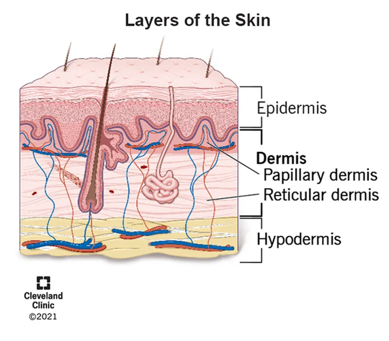
The skin is divided into 3 layers:
- Epidermis
- Dermis
- Hypodermis
Epidermis is the topmost layer of the skin that acts as a barrier to environmental pathogens and also contains melanocytes that regulate the amount of melanin and, thus, your skin color.
The dermis is divided into 2 parts (and this is important in burn depth classification):
Papillary dermis and reticular dermis, typically in a ratio of 20:80.
The papillary portion is the upper thinner part of the dermis that contains blood vessels, and the reticular is the lower thicker part with nerve endings, hair follicles, sweat, and sebaceous glands.
The hypodermis is the layer that contains subcutaneous fat, which also has hair follicles, blood vessels, and nerves.
What are the different degrees of burn?
There are 4 degrees of burns. Depending on the specific burns classification, there may be subdivisions, but all burns classifications will typically encompass all these 4 degrees.
As the depth of the burn increases, so does the degree of the burn.
First Degree
Involvement of only the epidermis. A typical example is a sunburn.
Second Degree
Superficial – involvement of epidermis and papillary dermis.
Deep -involvement of epidermis, papillary dermis, and only a part of the reticular dermis.
Third Degree
Involvement of the full thickness of skin (epidermis, papillar dermis, all of the reticular dermis, hypodermis)
Fourth Degree
Full thickness of the skin + underlying muscle and bone.
This severe degree of burn is typically seen in electrical contact burns and cases in which there has been prolonged contact with direct flame.
*Some classification systems will limit the fourth degree to only fat and classify involvement of the muscle and bone as fifth and sixth degree, respectively.
Salient features for burn assessment
- Burn depth is best assessed after 24 hours of the injury as the burn injuries are dynamic and may evolve over time to become deeper.
- How deep the burn wound is, or burns classification, is determined by the layers of the skin involved.
- Burns are almost never uniform in depth or degree. The same area of a burn can show areas with both superficial and deep burns.
- Improper first aid may result in a deepening of the burn depth, and thus it is essential to know the basics.
The Right Way To Give Burn First Aid At Home. - The burn depth determines the outcome- aesthetic or functional, making it essential to seek help from a trained professional.
Also read: How To Reduce Burn Scar? Top 10 Ways To Reduce Burns Scar At Home. - Certain types of burns can be serious and need immediate medical attention. E.g., burns on the hand, face, and genitalia, burns in children, the elderly, or those with disabilities and comorbid conditions like diabetes.
First degree burn
Sensitive Content
This photo contains sesitive content which some people may find offensive or disturbing

| Burns | Skin layer involved | Features | Healing time | Scarring potential |
|---|---|---|---|---|
| First Degree | Epidermis | Only redness with dry skin. Painful. Similar to a sunburn | < 5days | No raised scars. The burn area can darken if care is not taken. |
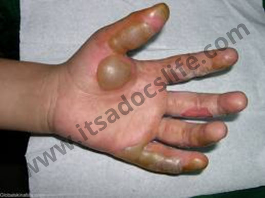
Second degree, superficial burn
Sensitive Content
This photo contains sesitive content which some people may find offensive or disturbing
| Burns | Skin layer involved | Features | Healing time | Scarring potential |
|---|---|---|---|---|
| Second Degree, Superficial | Epidermis & papillary dermis. | Extremely painful, blistering. The burn is wet, oozing, and pink-colored. | 10 to 21 days. | No raised scars but till new skin forms, there will be a wound. |
Second degree, deep burn
Sensitive Content
This photo contains sesitive content which some people may find offensive or disturbing
| Burns | Skin layer involved | Features | Healing time | Scarring potential |
|---|---|---|---|---|
| Second Degree, Deep | Epidermis, papillary dermis & only a part reticular dermis. | Less painful, may have blisters. The burn area has a white, waxy look with a mottled appearance and poor or no blanching when the area is pressed. | 4 to 6 weeks. | Potential for severe scarring that can lead to aesthetic and functional impairment. |
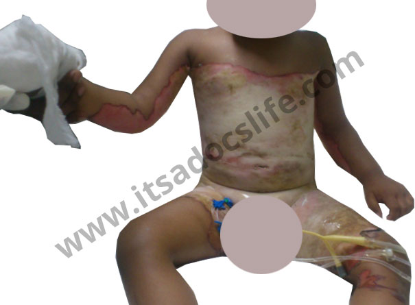
Third degree burn
Sensitive Content
This photo contains sesitive content which some people may find offensive or disturbing
| Burns | Skin layer involved | Features | Healing time | Scarring potential |
|---|---|---|---|---|
| Third Degree | Full thickness of the skin. | Painless due to nerve destruction. Skin is parchment-like or charred with visible thrombosed vessels. | Will require surgical intervention like skin grafting for healing. | Severe scarring leads to aesthetic and functional impairment. |
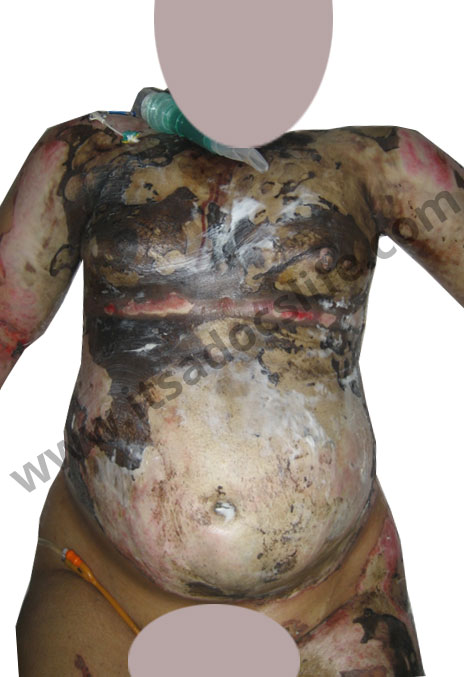
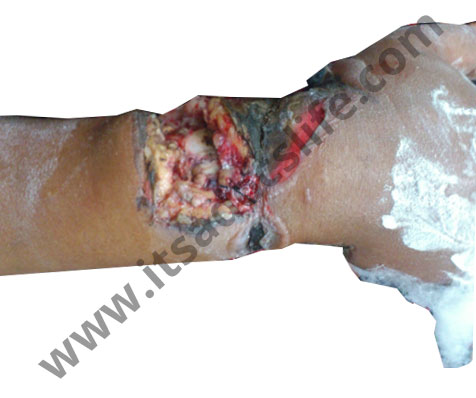
Fourth degree burn
Sensitive Content
This photo contains sesitive content which some people may find offensive or disturbing
| Burns | Skin layer involved | Features | Healing time | Scarring potential |
|---|---|---|---|---|
| Fourth Degree | Full thickness of skin + subcutaneous fat + muscle + bone with charring of limb or gangrene | Thick eschar to gangrene. No sensation in the area. | Life-threatening and may require amputation of the involved limb. | Often results in permanent disability and may require lengthy rehabilitation. |
*Some classification systems will limit the fourth degree to only fat and classify involvement of the muscle and bone as fifth and sixth degree, respectively.
Trivia
Clinical judgment by a trained professional remains the mainstay of assessment of a burn wound depth. Studies, however, have shown this to be accurate in only 60-75% of the cases, even when carried out by an experienced burn surgeon.